Testing of Glaucoma
Intraocular Pressure check
A patient’s intraocular pressure can be obtained with three different instruments: Tonopen, I-care and Goldman Applanation Tonometry. The “gold standard” and most accurate way of measuring your intraocular pressure is Goldman Applanation tonometry. This is achieved by placing a drop of fluorescent numbing solution into the eyes, placing the patient’s head in the slit lamp machine and shining a Cobalt blue light until the IOP is obtained by direct applanation.
For patients that have certain corneal surface irregularities or have small eyes with deep set orbits making applanation difficult, the Tonopen and I-care systems are usually a preferred way to measure the IOP.
Gonioscopy
Gonioscopy is a painless exam that is used to check a part of your eye called the drainage angle. It requires using a goniolens (a type of contact lens) as well as the slit lamp microscope to visualize the iridocorneal angle (the angle formed between the cornea and iris) which is where the fluid naturally drains out of the eye.
Gonioscopy is crucial in helping distinguish between the different types and causes of glaucoma.
Visual Field Testing
A visual field is used to test how wide of an area your eye can see when fixating on a central point. It is used to detect central or peripheral visual field loss that can be associated with glaucoma as well as multiple other causes.
This test is very important and is used to diagnose, stage, as well as monitor for disease progression of glaucoma. It is often obtained at least once yearly, sometimes more frequently if needed.
OCT and RNFL Analysis
Optical Coherence Tomography (OCT) is a non-contact imaging technique that allows measurement of the retinal nerve fiber layer (RNFL), a layer that is often thinned out in patients with glaucoma.
Because this structural change (loss/thinning of RNFL) often precedes visual field changes, OCT can improve a physician’s ability to detect early glaucoma and allow for timely intervention to prevent vision loss. OCT and RNFL analysis is also used in monitoring for progression of glaucoma, and is therefore obtained yearly.
Optic Nerve Photos
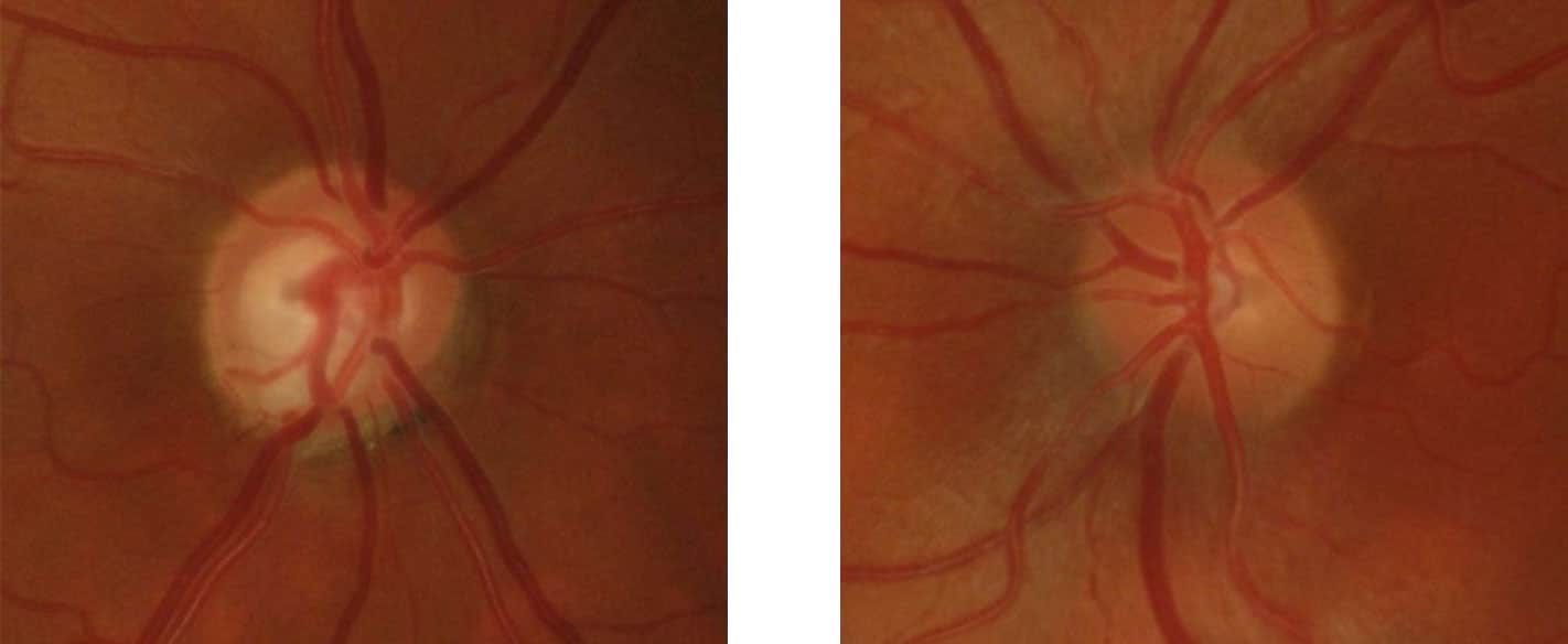
Optic Nerve Photos are used to detect and monitor for thinning of the neuroretinal rim tissue, which is another way of quantifying the degree of glaucomatous optic nerve damage and severity of the disease.
Photos are usually obtained every 2-3 years as a way of monitoring disease progression. They can also be used to document certain findings such as disc hemorrhages which can have important prognostic significance.

