Laser Glaucoma Treatment
Performed in clinic/office
SLT - Selective Laser Trabeculoplasty
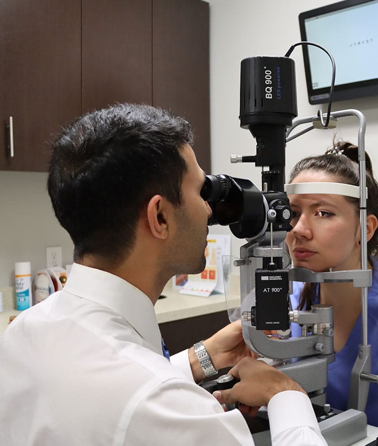
- Doctors perform Selective Laser Trabeculoplasty (SLT) as an outpatient procedure to reduce eye pressure in open-angle glaucoma patients. This non-invasive laser treatment happens under a microscope, much like the one used during your eye exams.
- Before starting, a doctor numbs your eye with drops and places a contact lens on it. They then aim the laser at the pigmented trabecular meshwork, which helps drain fluid from your eye, to enhance outflow and lower pressure. After the laser treatment, you’ll wait for a bit to get your eye pressure checked. You might experience blurry vision, slight discomfort, or light sensitivity for a few days. Doctors prescribe anti-inflammatory drops for a few days, and you’ll return for a follow-up in 4-6 weeks when the laser fully kicks in. SLT is a reliable method to manage ocular hypertension and open-angle glaucoma, delaying the need for surgery and can be repeated if necessary.
LPI - Laser Peripheral Iridotomy
- Laser Peripheral Iridotomy (LPI) is for patients with narrow angles or closed-angle glaucoma, where the drainage angle is mostly closed, causing high eye pressure, optic nerve damage, and potential vision loss. This condition can develop slowly (chronic) or suddenly (acute).
- LPI creates a tiny hole in the iris’s outer edge to open the drainage angle and improve fluid outflow. The procedure starts with drops to constrict the pupil and stretch the iris. After numbing your eye with drops, a contact lens is placed, and the laser makes a small hole in your peripheral iris. It typically lasts 2-5 minutes. You may feel minor pain or discomfort. Post-laser, you’ll wait to have your pressure checked. Doctors prescribe anti-inflammatory drops for a few days post-procedure. Some patients report temporary blurry vision, eye redness, light sensitivity, or discomfort, which generally improves within days. LPI effectively opens the drainage angle, ensuring proper fluid outflow.
Getting Ready for the Laser Glaucoma Treatment
Before your visit, collecting any previous medical records or details about your condition can be very helpful. This helps our doctors get a full picture of your health history.
The Glaucoma Center of Texas is more than a clinic—it’s a support network of caregivers, medical experts, and people like you, all working towards better vision and life quality.
Contact us now. Let’s start this journey to improved eye health together.

